 VENTRICULAR BIGEMINY
 The irregularity of the R–R interval is frequent with atrial fibrillation, nevertheless it should be noted that with a really fast HR during atrial fibrillation, the R–R interval could turn into common. Ventricular enlargement may alter QRS deflections, length or amplitude. Aberrant conduction, corresponding to through right or left bundle department, also can alter the QRS length and morphology (shape) for a similar cause. If conduction through a ventricular bundle department is blocked, the depolarization wavefront can't unfold along the fast conduction pathway within the affected ventricle.
Atrioventricular (AV) block; leads II and III, 25 mm/sec, 10 mm/mv. This electrocardiogram tracing signifies each second and third diploma AV block. The starting of the tracing exhibits some usually performed P waves making a sinus beat (SB). The P–Q interval is mounted at 80 ms, making this a Mobitz II second diploma AV block. The heart rate (HR) on the second-degree AV block in the beginning of the hint is roughly 57–60 bpm.
Previously, he spent 18 years at the University of Missouri in the cardiology service and 3.5 years at Ross University School of Veterinary Medicine in the anesthesia service. He presents internationally on veterinary cardiology topics. He has a passion for instructing, is the editor of Cardiology for Veterinary Technicians and Nurses, and has written over a dozen peer-reviewed articles. Areas of artifact should be recognized because the ECG is being recorded so they can be addressed if potential. Electric (60-cycle) noise may be due to poor electrical grounding (of the topic, the electrocardiograph or the table on which the ECG is being performed) or adjoining gear corresponding to lights or different electrical gear. Electric noise appears on the ECG as common fine, sharp, vertical oscillations. As talked about earlier, putting a hand on the animal’s thorax could help in reducing trembling or respiratory artifacts.
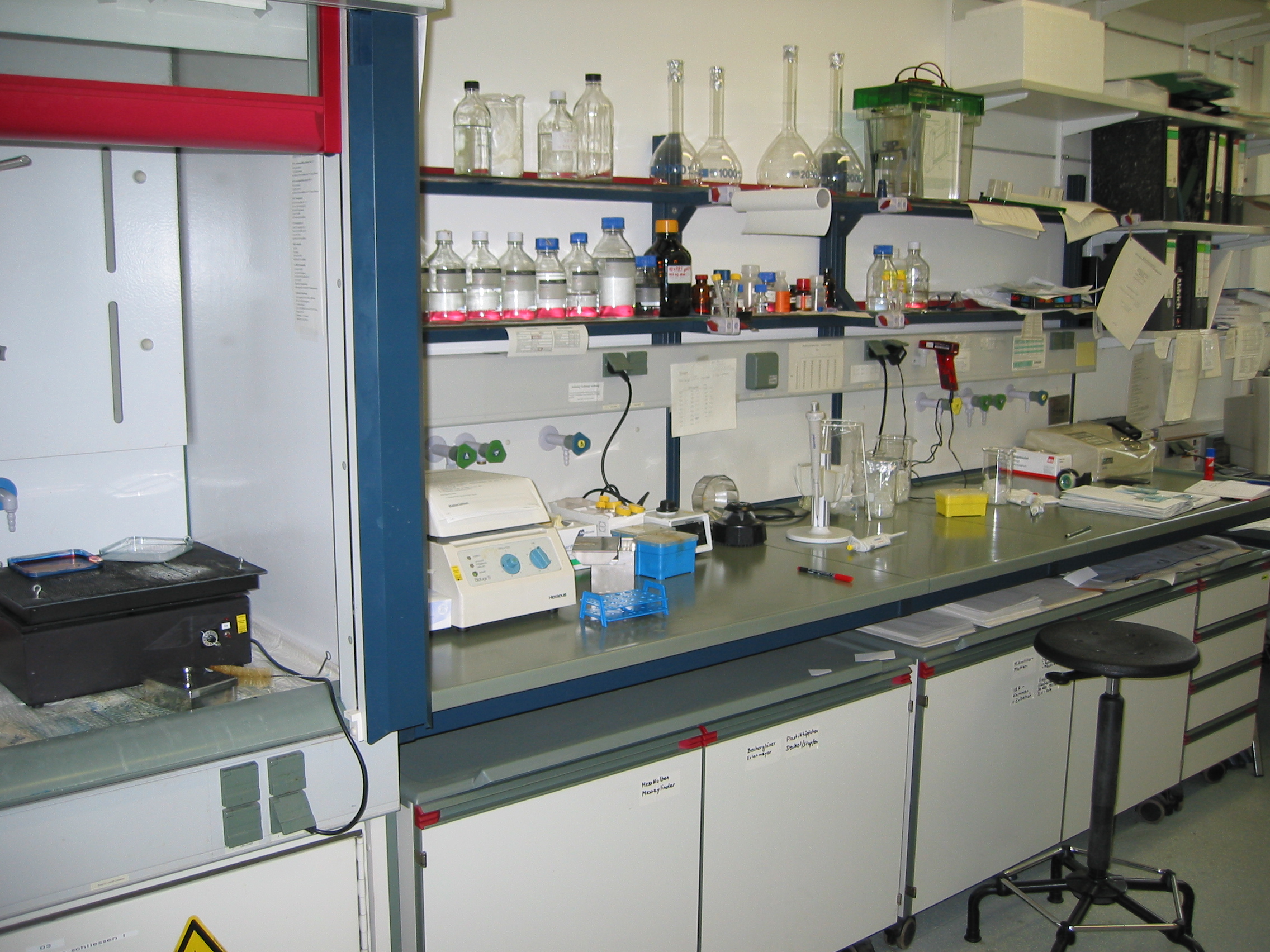
They also present persevering with training to the veterinary neighborhood each domestically and nationally. Echocardiography is the art of using ultrasound to view the construction and function of the guts in real time. Ultrasound is a extremely informative, non-invasive, and secure diagnostic test in both human and veterinary drugs. This technique makes use of excessive frequency sound waves emitted from a hand-held probe to supply an ultrasound beam. This ultrasound beam is mirrored from the tissues in the chest and heart and returns to the ultrasound probe to assemble an image of the center in movement.
Small Animal Ultrasound & Echocardiogram
How ECG monitoring contributes to patient care
Your renewal will assure uninterrupted service till the brand new expiration date of your renewed subscription. If the consumer would like to cancel their renewal, they have to accomplish that inside their account dashboard. Please evaluation our assist middle for extra data. VETgirl might provide alternatives for person interplay inside its Sites and social media profiles on sites such as Facebook, Twitter, LinkedIn, and numerous blogging websites.
Si seguimos teniendo dudas es aconsejable recurrir a algún servicio de telemedicina donde nos tienen la posibilidad de contribuir a interpretar nuestro electrocardiograma. El electrocardiograma debe efectuarse en una región sosegada dentro de la clínica, con el animal puesto en decúbito del costado derecho (a menos que haya alguna contraindicación para esto), y los miembros perpendiculares al tronco, intentado que el perro esté lo mucho más relajado viable a lo largo del examen. Comunmente los electrodos se colocan en los integrantes (proximal o distal al codo, pero teniendo en cuenta que la colocación proximal incrementa los artefactos, y sobre la rodilla), siguiendo el código de colores (rojo, previo derecho; amarillo, anterior izquierdo; verde, posterior izquierdo; negro posterior derecho). En Cediagvet contamos con un equipo de expertos con gran experiencia en el campo de la ecocardiografía veterinaria. Nuestro equipo está enormemente preparado y posee una amplia y extensa experiencia en la realización de estudios ecocardiográficos en animales. Puedes tener la tranquilidad de que tu mascota va a estar en las mejores manos cuando se someta a un examen ecocardiográfico en nuestro centro.
Fallo cardiaco agudo en el perro
En la actualidad, la percusión torácica solamente se efectúa en la clínica de pequeños animales debido a la extensa disponibilidad de la ecografía point of care (POCUS), pero se trata de una evaluación fácil que siempre es recomendable para identificar la línea horizontal de matidez socia al derrame pleural. Los parámetros más importantes que se deben controlar en el tratamiento del fallo cardiaco agudo son la continuidad y el ahínco respiratorios, así como la vitalidad general del perro. El fallo cardiaco agudo en el perro implica el peligro de muerte, con lo que, tal y como señala Luca Ferasin, es esencial optimizar el diagnóstico y el régimen. El líquido en la cavidad torácica y abdominal es visible tanto en una radiografía como en una ecografía.
Presentación clínica
En la mayoría de los casos, no se necesita sedación o anestesia para realizar el estudio, si bien en algunos casos específicos puede ser preciso sedar al animal para conseguir imágenes de gran calidad. LOS SITIOS, INCLUYENDO LOS SERVICIOS Y PRODUCTOS EN LOS SITIOS, SE PROPORCIONAN "TAL CUAL" Y "SEGÚN DISPONIBILIDAD" Y SIN NINGUNA GARANTÍA, EXPRESA O IMPLÍCITA. SU USO DE LOS SITIOS, INCLUIDOS SUS SERVICIOS Y PRODUCTOS, ES BAJO SU PROPIO RIESGO. VETGIRL RENUNCIA A TODAS LAS GARANTÍAS respecto a la precisión, seguridad y / o integridad de la información contenida EN LOS SITIOS. EN LA MEDIDA MÁXIMA PERMITIDA POR LA LEY APLICABLE, VETGIRL RECHAZA CUALQUIER REPRESENTACIÓN Y GARANTÍA DE CUALQUIER TIPO, EXPRESA O IMPLÍCITA, INCLUYENDO, PERO NO LIMITADO A, LAS GARANTÍAS IMPLÍCITAS DE COMERCIABILIDAD Y ADECUACIÓN PARA UNA PARTICULARIDAD.
Ecocardiografía en veterinaria
Nosotros aconsejamos aun realizar una investigación antes que haya sintomatología manifiesta (tos, ahogos, desmayos, insomnio, cansancio) ya que la medicación en la mayor parte de las ocasiones retrasa la evolución de la dolencia cardiaca pero no la cura. La radiografía del corazón nos permite comprender el tamaño del corazón y sus cámaras (atrios y ventrículos), laboratóRio de veterináRia esta manera cómo de los enormes vasos (aorta, cavas, y vasos pulmonares). Además nos permite conocer si los pulmones están encharcados de líquido y su alcance. El cardiólogo acostumbra realizar ecocardiograma o electrocardiograma si el gato o perro ha dado muestras de soplos u otros signos de patología cardíaca, tales como debilidad o adversidades respiratorias. VETgirl se reserva el derecho, cualquier ocasión, de cambiar o enmendar los Términos y Condiciones sin previo aviso. Las ediciones y / o enmiendas entran en vigencia justo después de su publicación en los Sitios.
"Qué hacer y qué no hacer" para el uso de furosemida en perros